Warsaw’s pioneering fetal spina bifida surgery
Warsaw: pioneering fetal spina bifida surgery
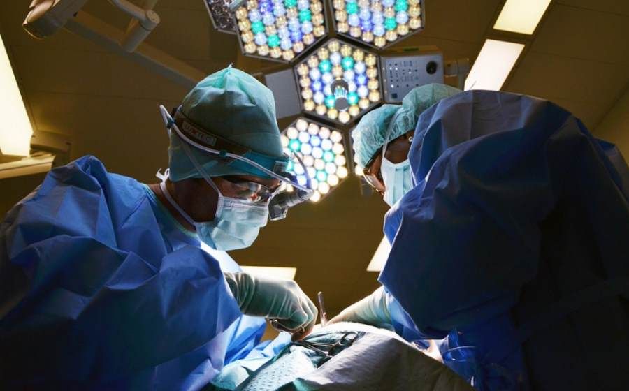
Poland’s pioneering fetoscopic surgery for spina bifida in a 25-week-old fetus was carried out by specialists from the Warsaw Medical University. The operation took more than four hours. Mom and toddler feel comfortable.
Spina bifida in a child is a defect in the continuity of the spine, most often in the lumbar or sacral region, which can lead to damage to the spinal cord. "The consequences of the defect are on the ogoł paralysis nog and paralysis of the sphincters of the bladder and/or rectum. Intrauterine surgery for spina bifida improves the prognosis for children diagnosed with the defect, reduces the risk of complications at birth, and offers a better chance of normal functioning of the lower limbs and sphincters” – explained in the WUM announcement.
The groundbreaking operation was carried out by the teamoł of the First Chair and Department of Gynecology and Obstetrics of the University of Medical Sciences (WUM), headed by prof. Mirosław Wielgosia: Dr. Przemysław Kosiński, Dr. Robert Brawura-Biskupski-Samaha, lek. Rafał Wojdacz, M.D., Monika Spain, and Sabrina Wagner and Prof. Thomas Kohl – precursor to fetoscopic fetal spina bifida surgery.
It is under the guidance of prof. Kohl’s University Hospital in Giessen, Germany, trained Polish specialists. The initiator and originator of the introduction of fetoscopic spina bifida surgery at the University Center for Women’s and Newborn Health, and thus in Poland, is Professor. Wielgoś.
As Dr. Kosinski explained, fetoscopic surgery for fetal spina bifida is an alternative to traditional surgery that proceeds with the opening of the uterine cavity, just as during a cesarean section.
– The new type of surgery involves making 3 holes in the uterine cavity, as in laparoscopy, and using these holes toow insert trocars into the amniotic cavity. This method of operation is called in the literature "keyhole method" (key hole surgery) or by a minimally invasive method. Carbon dioxide is introduced into the uterine cavity while pumping out amniotic fluid, whichory is the natural environment of the fetus. In this wayob very good visibility is achieved, as row also the possibility of electrocoagulation – the operation can be performed by cutting out a piece of abnormal tissue. Technically, the operation is very difficult – for example, the needle, ktoThe suture is only 13 mm,” added specialist.
It took 1.5 years to train the team at the German Clinic in Giessen and raise funds for the purchase of equipment. The operation was made possible thanks to the financial support of the Polsat Foundation.