A mandible was printed in Rzeszow, which minimizes intraoperative complications
Rzeszow has printed a mandible that minimizes intraoperative complications
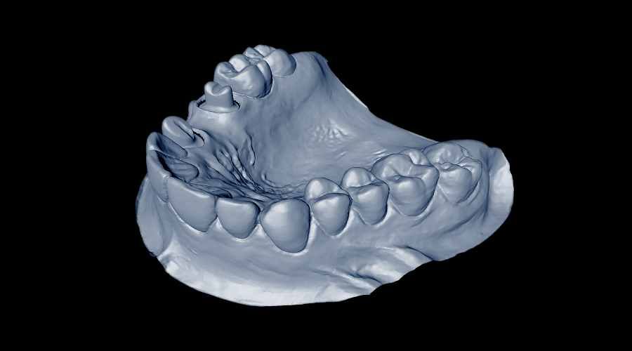
A model of a mandible made using 3D printing technology was created at the Rzeszow University of Technology. Its use significantly reduces the surgical time for implant placement and minimizes the occurrence of intraoperative complications.
The printed medical model of the mandible can be used by the surgeon to fit the implant to a particular patient, but even before the surgery. – It is tailor-made – mowi tworca model, Dr. Eng. Paweł Turek of the Rzeszów University of Technology. The research and the process of creating a model of the mandible became the subject of his doctoral dissertation titled “The process of creating a model of the mandible”. "Methodology for designing and manufacturing medical models of the mandible".
In an interview with PAP, Dr. Turek said that in the course of the research he consulted with specialists from the Department of Maxillofacial Surgery in Rzeszow, modified and refined the developed solutions, selected the appropriate parameters, until he reached a finished medical model of the mandible.
During his research, he was able to develop a procedure thatora allows computer reconstruction of the geometry of the mandible before the damage occurred, and to assess the degree of damage to the mandible. This means that the printed model is almost identical to the "original" anatomical mandible of a particular patient.
– TrojDimensional models of bone structures allow better preparation of the surgeon for the surgical procedure and increase precision, and this leads to significant reduction ofoThe system is designed to reduce surgery time and minimize the occurrence of complicationsopostoperative. In addition, the use of such a model helps reduce blood loss during surgery – mowired by Dr. Turek.
The printed model, whichory is individualized for each patient, allowing the surgeon to tailor an implant to a particular person, whichory to be implanted. This happens even before the surgery. In contrast, using the traditional procedure, the surgeon adjusts the, "covenants" implants into the patient during surgery, thus greatly extending surgery time.
In addition, the patient, after performing the procedure with the printed model, undergoes a faster recovery period and – according to the degree of damage to the mandible – can wrocic to function normally.
Another advantage of the troThe jdimensional model is that the patient, seeing it before the procedure, can more easily imagine the surgeon’s actions when the surgeon explains the entire process to him or her.
Tworca of a 3D-printed model of the mandible, he stipulated, however, that it was not a simple task to design and make an accurate medical model for the purpose of performing surgery to. The point is that the mandible is a bone tissue with a very complex geometry.
– The need for appropriate knowledge and skills in medicine and technical sciences, whichore allow to make full use of the tools to carry out such an operation – emphasized the scientist. He added that this is where the need for close support of theoThe surgeon is able to adjust the implant to a particular person, which allows him or her to work in cooperation and consultation with specialists.
As he explained, he chose the mandible because it is the only movable craniofacial bone and is subjected to multidirectional dynamic loads during biting or chewing.
The research process of Dr. The Turk was funded as part of the Innovation Incubator’s activities "Methodologies for designing and manufacturing medical models of the craniofacial with high accuracy".
The researcher intends to soon patent the model of the mandible he has developed.
He is currently conducting research on the cranial vault (the idea is to improve the process of its segmentation and reconstruction) and on the accurate fabrication of small and complex objectsow, such as. molars and dental crownsow.
– Reconstructive engineering is used today in many fields, such as in medicine, where it can be used, for example, as an. In the process of reconstructing the geometry of internal anatomical structures and manufacturing implantsow – noted Dr. Turek.
He is also conducting research within the Cluster Association "Technology in Medicine" – TECHNOMED, where it makes surgical models and templates using 3D printing techniques m.in. pelvis, femur, ktore significantlyob make it easier to plan and carry out a surgical procedure.
He assured that as part of the cluster’s activities, he plans to expand his research to other bone structures. – Their selectionor will depend on demand – noted
Sourceobackground: PAP – Science in Poland , Agnieszka Pipała, fot. JAKO5D/Pixabay